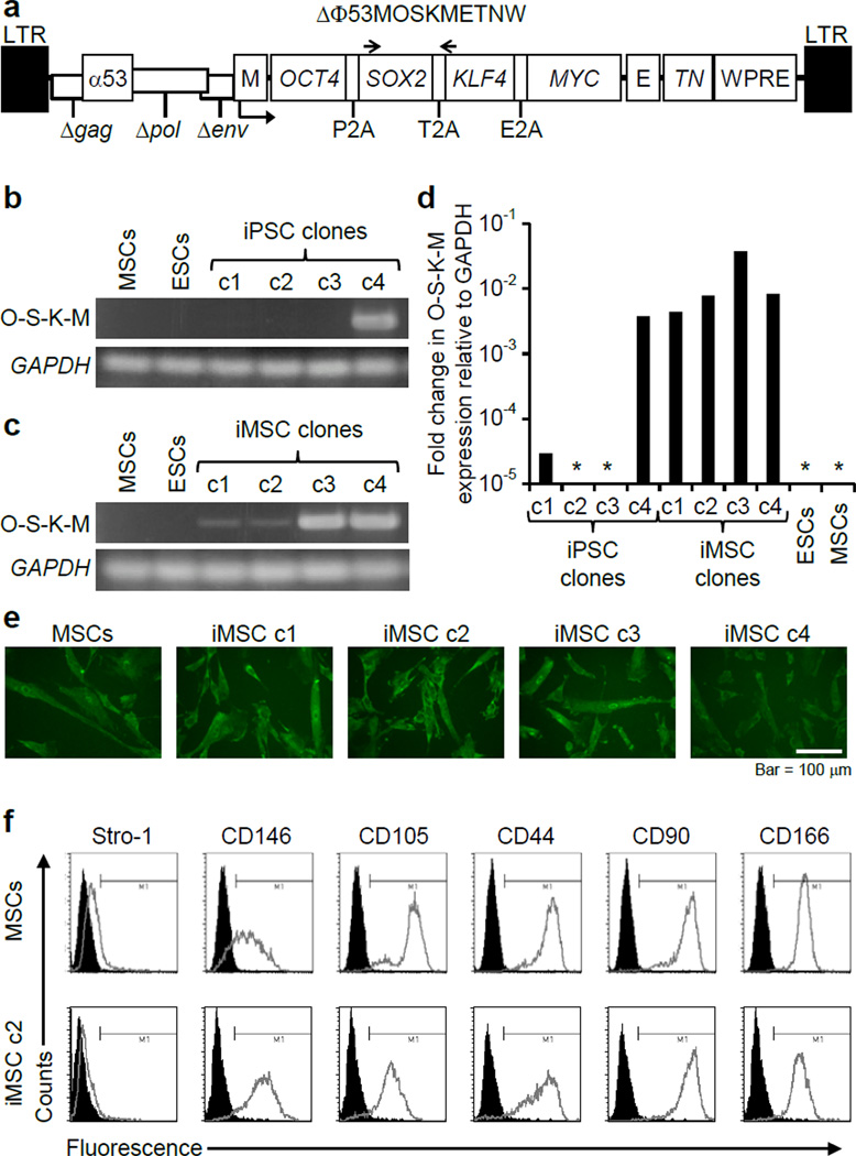
Figure 1. iPSC derivation and differentiation.
(a) The FV vector ΔΦ53MOSKMETNW is shown containing a polycistronic 2A peptide-linked reprogramming cassette with OCT4, SOX2, KLF4, and MYC open reading frames. E, EF1α promoter; M, MLV promoter; TN, Thymidine kinase-neomycin fusion protein gene; α53, anti-p53 shRNA; WPRE, woodchuck hepatitis virus posttranscriptional regulatory element. The locations of primers Foamy-f and Foamy-r are indicated by arrows. (b) RT-PCR analysis showed silencing of the FV polycistronic transcript in three of the four iPSC clones during reprogramming, with GAPDH transcript controls. O-S-K-M, reprogramming vector transcript. (c) RT-PCR showing expression of the reprogramming vector after differentiation of iPSCs into iMSCs. (d) mRNA levels of the FV polycistronic transcript (O-S-K-M) as determined by qRT-PCR and shown as fold change relative to GAPDH. *No transcript detected. (e) Collagen expression detected by immunohistochemistry with anti-human α2 Type I procollagen antibody in MSCs and iMSCs. Bar = 100 µm. (f) Representative flow cytometry analysis of MSC surface markers produced by MSCs and iMSC c2.

