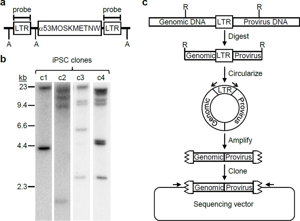
Figure 2. Identification of FV integration sites.
(a) Diagram of an integrated reprogramming vector with locations of the LTR probe shown. A, Avr II sites. (b) Southern blot analysis of Avr II-digested genomic DNAs to determine the number of FV vector integration sites in each iPSC clone. Each integrant produces 2 LTR-hybridizing fragments. (c) Inverse PCR strategy for identifying chromosome-provirus junctions. R, restriction enzymes sites; open arrows, LTR-specific PCR primers; jagged box, LTR remnant; closed arrow, sequencing primers.

