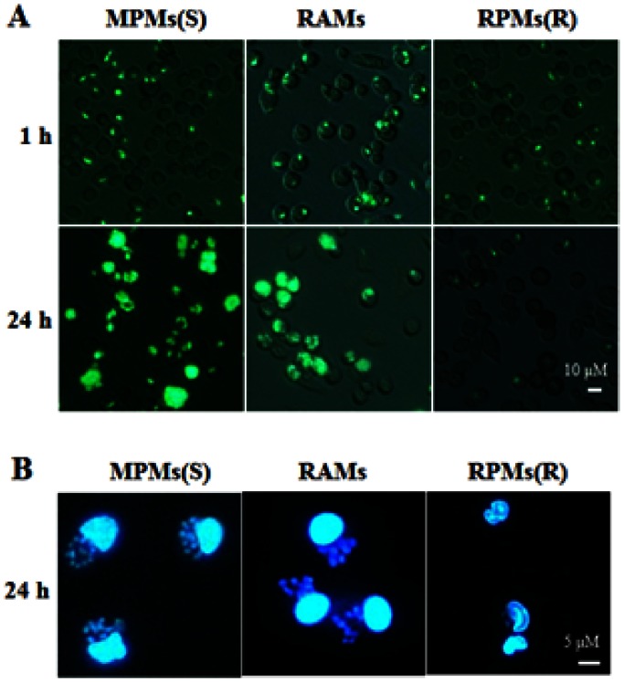
Figure 1. Analysis of T. gondii proliferation in rat alveolar macrophages in vitro.
Rat alveolar macrophages (RAMs) were incubated with T. gondii at the ratios of 1∶1 (parasites/macrophages = 1∶1), then the extracellular T. gondii (the non-invaded individuals) were washed from the medium and the time was defined as 1 hr. After 24 hrs, cells from the same cultures were compared. Mouse peritoneal macrophages (MPMs) (susceptible) and rat peritoneal macrophages (RPMs) (resistant) were designated as controls in the infection of T. gondii. (A and B) Different methods of Fluorescence Microscopy or DAPI staining were used in these experiments to observe the infection of T. gondii in macrophages. All the results were observed by fluorescence microscopy using a 350 nm exciting wavelength and 495 nm emitting wavelength. The results are representative of three similar experiments.

