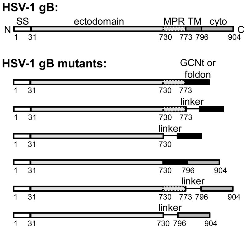
Fig. 1.
Schematic view of the sequence of the full-length HSV-1 gB and constructs designed to stabilize the prefusion conformation. SS – signal sequence, MP – membrane-proximal region, TM – transmembrane region, Cyto – cytoplasmic domain are shown as rectangles of different shades of gray. GCNt (sequence: QIEDKIEEILSKIYHIENEIARIKKLIGE) and foldon (sequence: GYIPEAPRDGQAYVRKDGEWVLLSTF) are shown as black rectangles. Linkers GSGS, GSGTGS, or GGSGGTGGSG are shown as lines.

