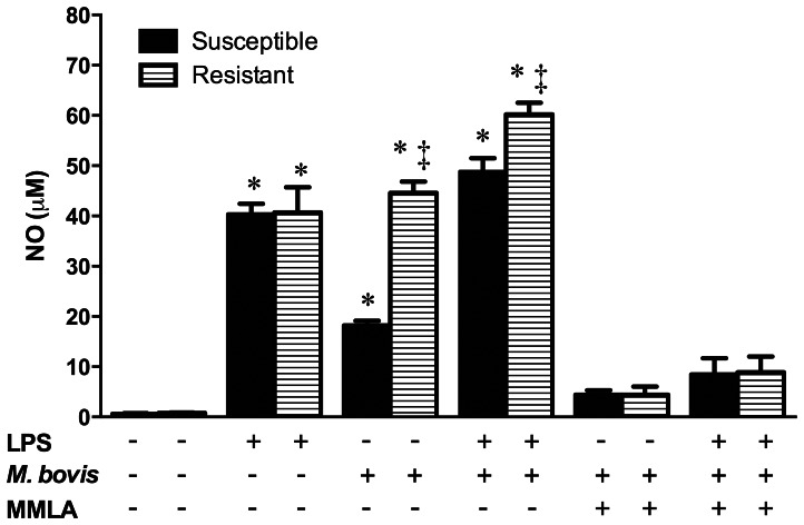
Figure 3. Mycobacterium bovis nitric oxide differential induction in susceptible and resistant bovine macrophages.
Macrophages stimulated or not with purified E. coli 026:B6 LPS (100 ng/mL) for 22 h, were infected with M. bovis field strain 129QP (MOI of 10) for 4 h. Cells were washed and cultured again for 24 h in presence or absence of the nitric oxide inhibitor n G-monomethyl-L-arginine monoacetate (MMLA) and nitric oxide was measured in cell culture supernatants by Griess assay. Results are mean ± standard deviation of quadruplicates of two independent experiments with macrophages of three susceptible and three resistant cows. Statistical differences (P<0.05) of each bar with its respective control (no treatment) (*) and among phenotypes on each condition (‡) are indicated.

