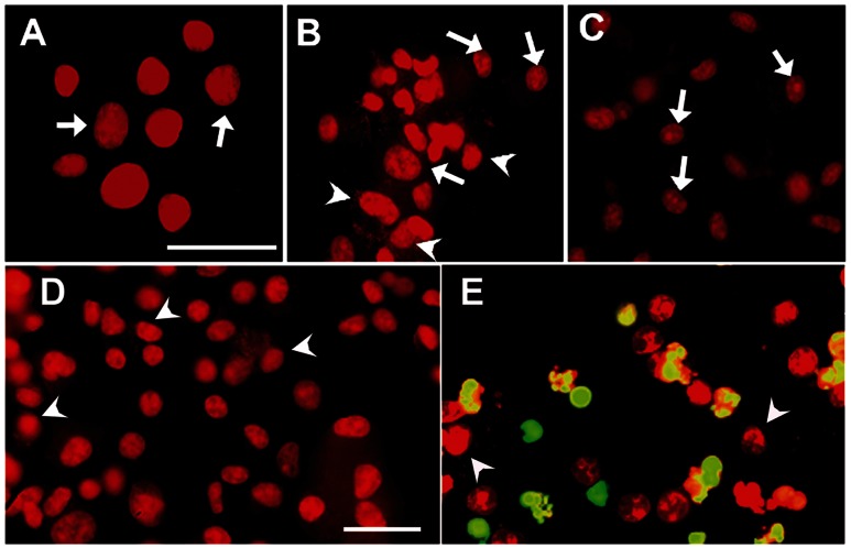
Figure 5. Apoptosis induction in Mycobacterium bovis-infected bovine macrophages.
Uninfected macrophages (A), macrophages treated with camptothecin (10 µg/mL for 48 h) (C) and macrophages infected with Mycobacterium bovis field strain 9926 (B, D and E), were washed at 4 h and cultured again for 24 h. Then cells were processed by TUNEL in presence (E) or absence (D) of TdT enzyme. Chromatin condensation was present in no-infected (arrows) and infected cells (arrowheads). TUNEL-positive cells show a green-yellow mark in panel E (BrdUTP-FITC). Magnification for A to C is 63X and for D and E is 40X with a scale bar of 50 µm each.

