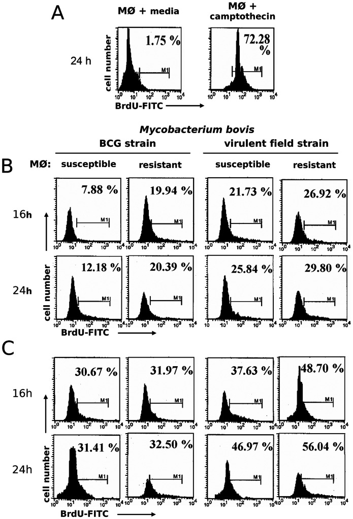
Figure 6. Mycobacterium bovis induction of bovine macrophage DNA fragmentation is time dependent.
Adherent-macrophages (MØ) were cultured with media alone or with camptothecin (10 µg/mL for 48 h) as negative and positive controls, respectively (A). Also macrophages were cultured in absence (B) or presence of purified E. coli 026:B6 LPS (100 ng/mL) for 22 h (C), and infected with BCG or M. bovis field strain 9926 (MOI of 10 for 4 h), washed and cultured again for 16 and 24 h and stained with TUNEL (BrdUTP-FITC). Histograms are frequency distributions of 1×10⁵ macrophages along FITC signal (log10 scale). Values are percentages of true BrdU-FITC TUNEL-positive cells (M1 gate) of one experiment, representative of two independent experiments with macrophages of three susceptible and three resistant cows. One-way ANOVA showed significant variation in infected versus uninfected controls (P<0.05) but not between phenotype (P = 0.4544) regardless of M. bovis strain.

