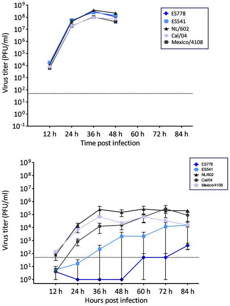
Figure 5. In vitro replication kinetics of A(H1N1)pdm09-like viruses isolated from Northern elephant seals showing growth curves of the two elephant seal pH1N1 isolates in Madin Darby Canine kidney (MDCK) cells (A) and Human Tracheobronchial Epithelial (HTBE) cells (B) compared to three reference stains: A/California/04/2009, A/Mexico/4108/2009, A/Netherlands/602/2009.
Supernantants were collected at the indicated time points and titrated by standard plaque assay, graphs show the mean titres of triplicate wells per time point and error bars indicate the standard deviation. The dotted line represents the limit of detection of 50 PFU/m.

