FIGURE 4.
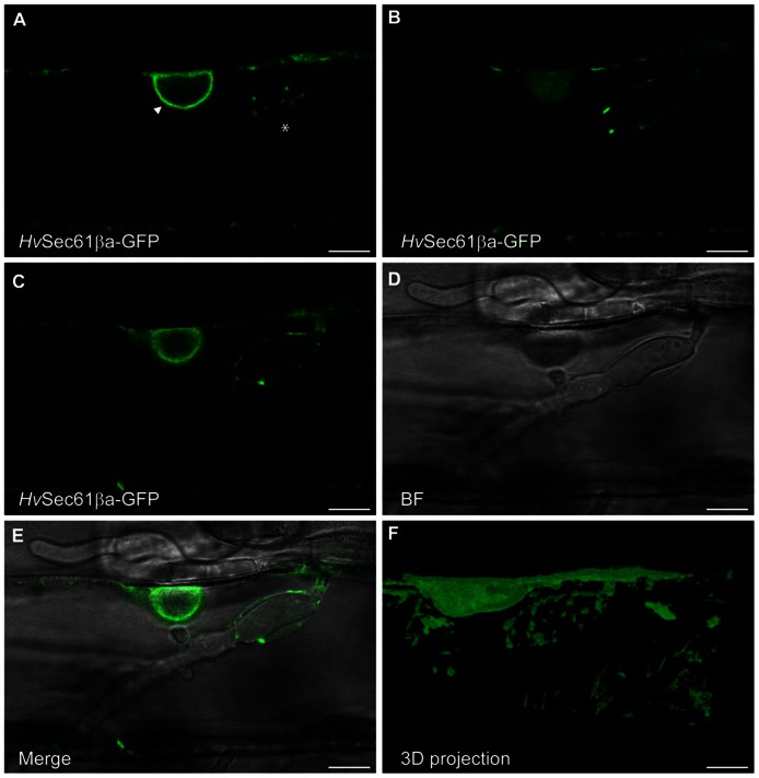
HvSec61βa-GFP localizes around the Bgh haustorial body. Confocal image of an epidermal cell, transformed with the HvSec61βa-GFP construct, taken 48 h after Bgh inoculation. (A–C) Three different focal planes from an image series of an infected cell with a haustorium. HvSec61βa-GFP localizes to the ER around the nucleus (arrow head, A) and surrounds the haustorium in an ER-like tubular pattern (asterisk, A). (C–E) GFP fluorescence (C), bright field (BF) (D) and merged image (E) show HvSec61βa-GFP localization at the surface of the haustorial body. HvSec61βa-GFP labels the tubular ER network, which is further illustrated in the 3D projection (F) (Image Surfer 1.2). Scale bar, 10 μm.

