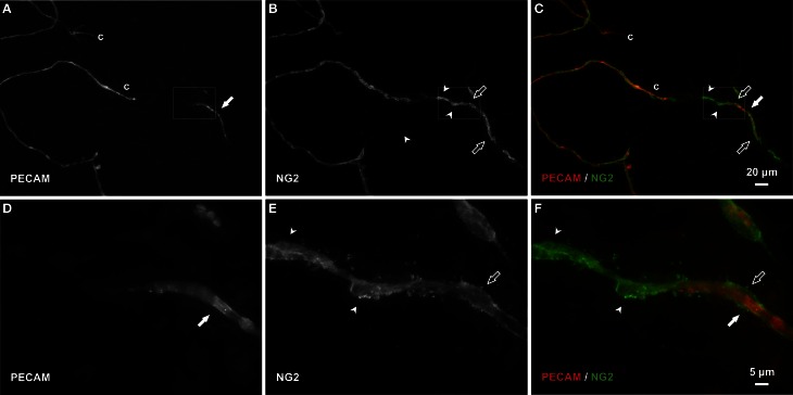
Figure 5.
Pericytes bridge and extend along vascular islands during regression. (A–C) Examples of PECAM-positive endothelial cells along vascular islands (filled arrows) with positive NG2 pericyte association. NG2 positive pericytes were observed associating with islands by either wrapping along (open arrows) or bridging (arrowheads) endothelial cells. (D–F) Higher magnification images of PECAM and NG2 labeling within the area defined by the rectangle. Blind ended capillary segments still connected to a network are indicated by “c.”

