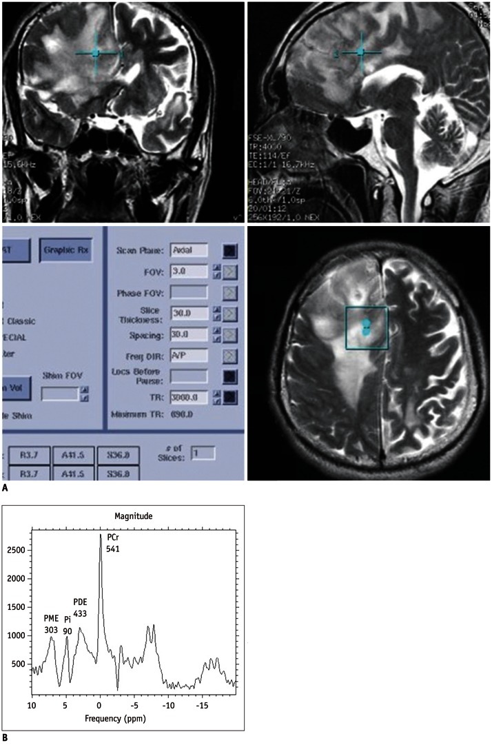
Fig. 3.
44-year-old man with high grade astrocytoma of brain.
A. Localizer image for 31P MRS shows huge mass in right frontal lobe without cyst or hemorrhage. B. 31P MRS shows high level of PME (303), low level of Pi (90) and relatively low level of PDE (433). Calculated PME/PDE ratio (0.70) is higher than its mean value in control group (0.47). PME = phosphomonoester, PDE = phosphodiester, PCr = phosphocreatine, MRS = magnetic resonance spectroscopy, Pi = inorganic phosphate

