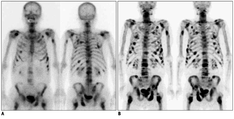
Fig. 1.
Findings of bone positron emission tomography (PET) and bone scan in 84-year-old male prostate cancer patient (no.2 in Table 1) with numerous bone metastatic lesions.
A. Anterior and posterior planar images of bone scan show multiple bone metastases. B. Bone PET anterior and posterior maximum-intensity projection images reveal numerous metasatic bone leisons. Please note that metastatic lesions on bone PET are more prominent than those on bone scan.

