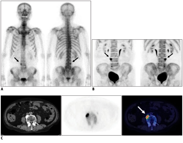
Fig. 2.
Case demonstration showing higher specificity of bone positron emission tomography (PET) than bone scan in 77-year-old male patient with colon cancer (no.14 in Table 1). He has been complaining of intractable back pain.
A. Bone scan shows focally increased uptake at L4 vertebral body (black arrows), which was suspected of bone metastasis. B. Bone PET maximum-intensity projection images also revealed hot uptake at same location (black arrows). C. Trans-axial images of CT, bone PET, and fusion image (from left to right) clearly demonstrate that osteophyte at L4 has intense uptake of Na18F (white arrow). It is noteworthy that tomographic images (C) play decisive role in determining nature of osteophyte.

