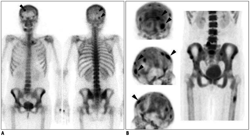
Fig. 3.
Case demonstration showing higher sensitivity of bone positron emission tomography (PET) than bone scan in 44-year-old female patient with breast cancer (no.3 in Table 1).
A. Anterior and posterior bone scan images show only 3 abnormal foci in skull (arrowheads). B. Skull lesions observed in bone scan are found to be more prominent in bone PET (arrowheads) (top, posterior maximum-intensity projection [MIP]; middle, right lateral MIP; and bottom, left lateral MIP images). In addition, many other bone metastatic lesions are found in skull, lumbar spines, sacrum, pelvic bones, and left femur.

