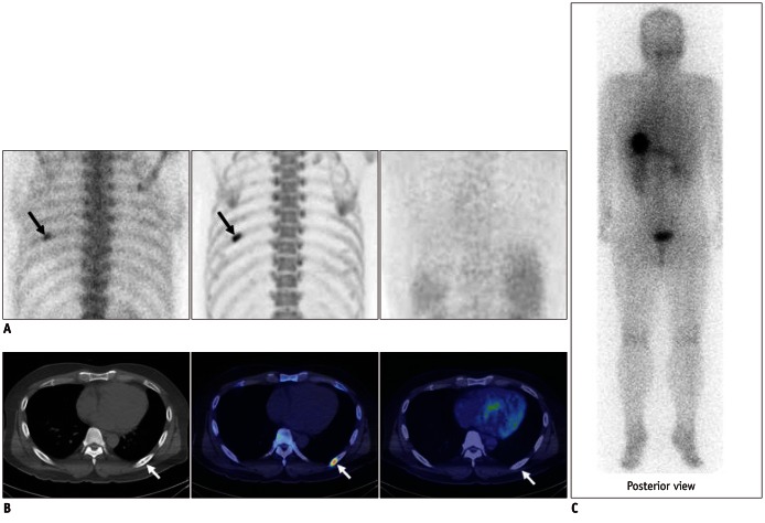
Fig. 4.
Case demonstration showing false positivity of bone positron emission tomography (PET) and bone scan in 39-year-old male patient with thyroid cancer (no.9 in Table 1).
A. (left, bone scan posterior planar; middle, bone PET posterior maximum-intensity projection [MIP]; and right, 18F-FDG PET posterior image) Bone scan and bone PET revealed abnormal uptake in left 7th rib posterior arc (black arrows); however, 18F-FDG PET was negative in left 7th rib area. B. (left, CT; middle, fusion of bone PET/CT; and right, fusion of 18F-FDG PET/CT) CT revealed osteosclerotic lesion in left 7th rib posterior arc, which was compatible with hot uptake in bone PET/CT; however, lesion was negative in 18F-FDG PET/CT (white arrows). C. Posterior image of 131I whole body scan obtained 2 days after administration of 131I (30 mCi). No lesion was found in left 7th rib area.

