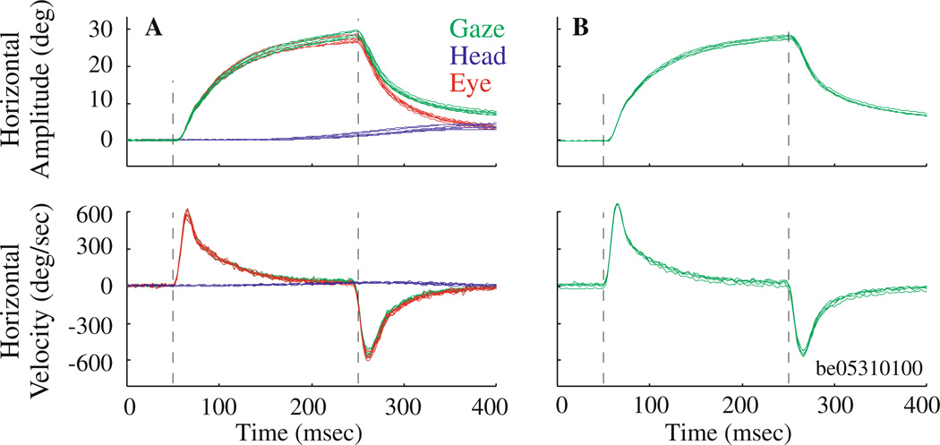
Fig. 8.
Microstimulation of the abducens nucleus in a head-unrestrained and b head-restrained conditions. a Top and bottom panels plot horizontal components of stimulation-evoked changes in position and velocity, respectively, in gaze (green), eye-in-head (red) and head-in-space (blue). b Same format at a, but only gaze profiles (green) are displayed; eye-in-head equals gaze because head-in-space is held constant at zero. Stimulation parameters: 25 µA, 200 ms, 300 Hz. Vertical dashed lines indicate stimulation onset and offset. These representative trials for both head-restrained (n = 5 trials) and head-unrestrained (n = 7 trials) data were evoked from the same site

