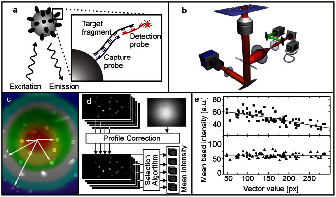
Figure 1. Principle of bead-based DNA quantification.
(a) Target DNA fragments are captured by beads functionalized with specific complementary oligonucleotides (capture probe) and labeled by reporter oligonucleotides with a fluorophore attached (detection probe). (b) Fluorescence of the beads is measured on a compact, robust and simplified wide-field fluorescence microscope. (c–e) Data analysis. (c) Beads are excited with different excitation intensities dependent on their position in the field of view. (d) Principles of data processing. The image is corrected for the variation in excitation across the image and then single beads are selected for analysis. (e) Dependency between mean bead intensity and position in the field of view.  Raw and
  profile corrected intensity values.

