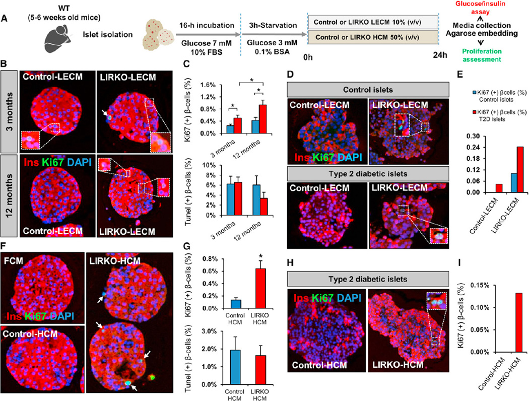
Figure 5. Hepatocyte-Derived Factors Stimulate Mouse and Human Islet β Cell Replication In Vitro.
Five to 6-week-old mouse islets were stimulated for 24 hr with LECM or HCM obtained from control or LIRKO mice. Islets were embedded in agarose and subsequently analyzed by immunostaining. Culture media were assayed for glucose and insulin.
(A) Schematic of the experimental protocol. See also Figure S6.
(B) Representative images of mouse islets stimulated with LECM derived from 3-month-old (upper panel) and 12-month-old animals (lower panel).
(C) Quantification of Ki67+ insulin+ cells (upper panel) and TUNEL+ insulin+ cells (lower panel): at least 3,000–5,000 cells were counted in each experimental group (n = 5 in each group).
(D) Representative images of healthy human donor islets (upper panel) and type 2 diabetic donor islets (lower panel) treated for 24 hr with LECM derived from control versus LIRKO mice. See also Table S3.
(E) Quantification of Ki67+ insulin+ cells in (D): between 3,000 and 4,000 cells were counted in each condition (Control LECM versus LIRKO LECM) in control islets, and at least 2,000 cells were counted in each experimental group for type 2 diabetic islets. See also Table S3.
(F) Representative images of mouse islets stimulated with HCM derived from 6-month-old control versus LIRKO mice or fibroblast-conditioned media (FCM).
(G) Quantification of Ki67+ insulin+ cells (upper panel) and TUNEL+ insulin+ cells (lower panel): between 4,000 and 5,000 cells were counted in each experimental group (Control HCM versus LIRKO HCM) (n = 5 in each group).
(H) Representative images of type 2 diabetic donor islets stimulated with control or LIRKO HCM. See also Table S3.
(I) Quantification of Ki67+ insulin+ cells in (H): at least 2,000 cells were counted in each experimental condition. See also Table S3.
Data represent mean ± SEM. *p ≤ 0.05. (See LECM Stimulation, HCM Stimulation, and Human Islet Studies sections in Experimental Procedures.)

