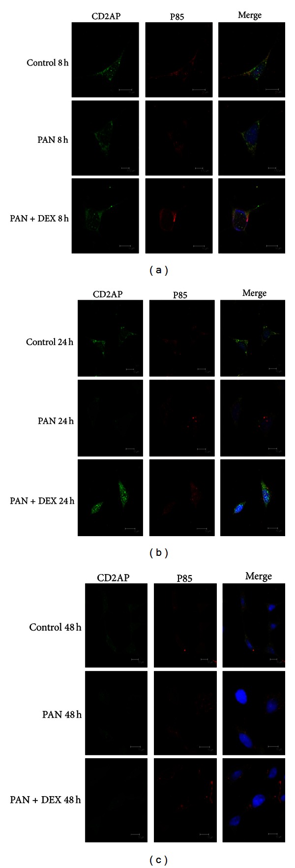
Figure 2.

Changes in the co-localization of CD2AP and p85 in control cultures, PAN-treated cultures and PAN + DEX co-treated cultures as revealed by immunocytochemistry under confocal laser scanning microscopy (600x). In control cultures, significant overlap between the green fluorescent staining of CD2AP and the red fluorescent staining of p85 was observed in the cytoplasm, plasmamembrane, and nucleus. PAN-treated cultures showed decreased overlap in the cytoplasm at 24 and 48 h, but enhanced co-staining of the nuclear envelope and nucleus. Reduced nuclear fluorescence overlap at 24 h and 48 h in PAN + DEX co-treated cells.
