Figure 2.
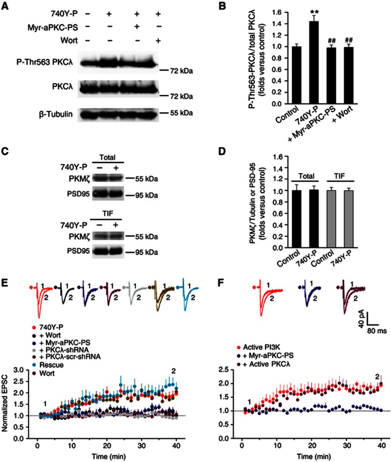
PKCλ functions downstream of PI3K to induce LTP. (A) PI3K selectively activated PKCλ. PI3K activation by 740Y-P treatment specifically activated PKCλ, indicated by potentiated PKCλ phosphorylation at Thr563 site. This activation of PKCλ was abolished by PKCλ antagonist Myr-aPKC-PS or PI3K antagonist Wortmannin. By contrast, total PKCλ was not affected by PI3K activation. (B) Plots summarize data from the experiments in (A) showing the Thr563P-PKCλ/total PKCλ ratio under various treatments (740Y-P, 1.44±0.11, n=8, compared with control, P<0.01; Myr-aPKC-PS, 0.98±0.05, n=8, compared with 740Y-P, P<0.01; Wort, 0.99±0.06, n=8, compared with 740Y-P, P<0.01; one-way ANOVA LSD test). **Compared with control, P<0.01; ##compared with 740Y-P, P<0.01. (C) 740Y-P treatment did not change the total amount of PKMζ or PKMζ amount at PSD. (D) Plots summarize data from the experiments in (C) showing absence of changes in the total PKMζ/PSD95 (740Y-P, 1.01±0.07, n=7; compared with control, P>0.05, independent samples t test) or TIF PKMζ/PSD95 ratio under 740Y-P treatment (740Y-P, 1.00±0.04, n=4; compared with control, P>0.05). (E) PKCλ is required for LTP induced by PI3k activation. Activation of PI3K with PI3K agonist 740Y-P (15 μM) induced long-lasting potentiation of EPSCs (1.87±0.21, n=6; compared with baseline, P<0.05, paired sample t test). 740Y-P was applied in the recording pipette solution. This enhancement of AMPA EPSCs was significantly attenuated by blocking PKCλ with Myr-aPKC-PS (2.0 μM; 1.14±0.11, n=6; compared with 740Y-P, P<0.01, one-way ANOVA LSD test) or by selective PI3K antagonist Wort (0.5 μM; 1.15±0.13, n=6; compared with 740Y-P, P<0.01). As a control, Wort alone failed to exert any effect on basal EPSCs (0.97±0.13, n=7; compared with baseline, P>0.05). PKCλ knockdown attenuated enhancement of AMPA EPSCs (0.92±0.06, n=7; compared with baseline, P>0.05; compared with 740Y-P, P<0.01), which was reversed by PKCλ rescue (2.25±0.27, n=5; compared with 740Y-P, P>0.05; compared with PKCλ KD, P<0.01). In contrast, LTP was normal in cells transfected with PKCλ-scr-shRNA (1.79±0.16, n=5; compared with 740Y-P, P>0.05; compared with PKCλ KD, P<0.01). (F) PI3K-induced LTP occludes further enhancement induced by active PKCλ. Active PI3K (2 μg/ml), when applied in pipette solution, induced persistent enhancement of EPSCs (1.84±0.14, n=6; compared with baseline, P<0.01). This form of LTP was abolished by blocking PKCλ with Myr-aPKC-PS (2.0 μM; 1.08±0.08, n=6; compared with active PI3K, P<0.01). When co-applied with active form of PKCλ (1.5 nM), however, PI3K-induced LTP failed to display further enhancement in the magnitude of LTP (1.78±0.22, n=6; compared with active PI3K, P>0.05).
Source data for this figure is available on the online supplementary information page.

