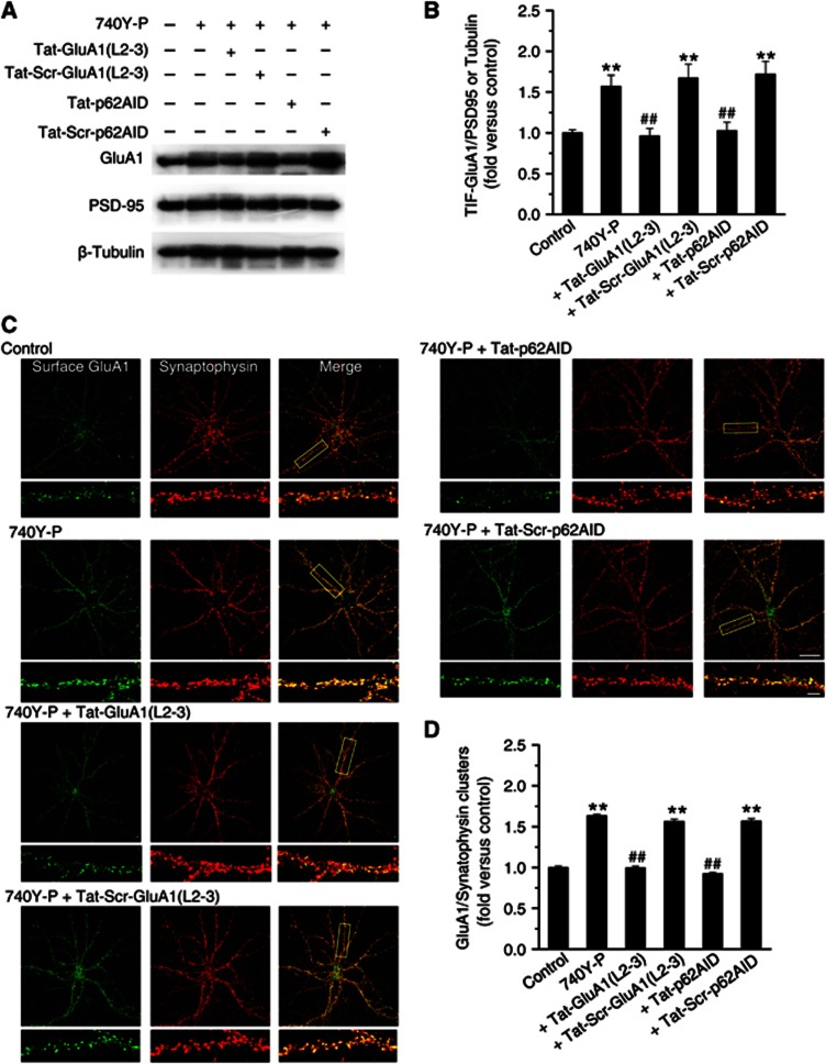
Figure 6.
Association of PKCλ or GluA1 with p62 is required for PI3K-induced AMPAR insertion at synapses. (A) Interfering peptides affected PI3K-induced increase in GluA1 expression at postsynaptic sites. (B) Plot summarized the data from experiments shown in (A). Increase in GluA1 postsynaptic clustering upon PI3K activation was completely reversed by interfering GluA1/p62 association with Tat-GluA1(L2-3) or by interfering p62/PKCλ association with Tat-p62AID (740-Y-P+Tat-GluA1(L2-3), 0.96±0.09, n=8, compared with 740-Y-P, 1.57±0.14, n=8, P<0.01; 740Y-P+Tat-p62AID, 1.03±0.10, n=8, compared with 740-Y-P, P<0.01; one-way ANOVA LSD test). The scrambled peptides (Tat-Scr-GluA1(L2-3) and Tat-Scr-p62AID) failed to display effect in GluA1 postsynaptic expression (compared with 740-Y-P, P>0.05). (C) Interfering peptides suppressed increase in AMAPR insertion upon PI3K activation. Example images of cell surface AMPARs (green; non-permeabilized) at synapses (red; synaptophysin) under control and various treatments are shown. Bar, 30 μm. Higher magnification of dendritic branches are in lower panels. Bar, 5 μm. The boxes in the Merge column indicated corresponding magnified regions. (D) Plot summarized the data from experiments shown in (C). Both of the two interfering peptides reversed increase in surface AMPAR insertion at synapses (740-Y-P+Tat-GluA1(L2-3), 1.00±0.02, n=19, compared with 740-Y-P, 1.63±0.03, n=22, P<0.01; 740Y-P+Tat-p62AID, 0.92±0.02, n=20, compared with 740-Y-P, P<0.01). As a control, the scrambled peptides failed to display any significant effect on AMPAR insertion (P>0.05). **Compared with control, P<0.01; ##compared with 740Y-P, P<0.01.
Source data for this figure is available on the online supplementary information page.

