Figure 7.
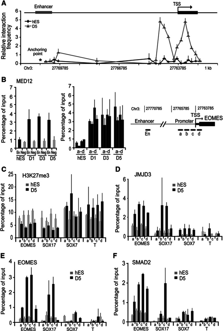
The sequential two-step activation of EOMES locus is conserved between human and mouse. (A) The quantitative 3C analyses on Eomes locus was carried out in hES cells and definitive endoderm derived from hES cells (D5). Similar to mES cells, enhancer–promoter looping is absent in undifferentiated hES cells but accompanies EOMES gene activation in differentiated cells. (B) Using ChIP analyses, MED12 bound to both enhancer (En) and promoter (a–d) of the EOMES locus but not to the negative control region (Neg), confirming looping formation in Activin A-differentiated hES cells. (C–F) EOMES activates selectively definitive endoderm loci in Activin A-differentiated hES. ChIP analyses on definitive endoderm derived from hES cells (D5) that H3K27me3 (C) is reduced and JMJD3 (D), EOMES (E), and SMAD2 (F) are enriched at the promoter regions (a–d) of EOMES and SOX17, but not SOX7 and BRA. Values are mean±s.e.m. (n=3–4).

