Figure 1.
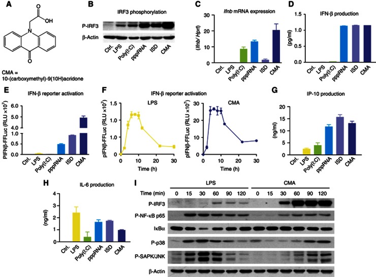
CMA strongly induces type I IFN in primary mouse macrophages. (A) The chemical structure of CMA is depicted. (B–E and G–H) Bone marrow-derived macrophages were transfected with poly(I:C), pppRNA and ISD, or stimulated with LPS or CMA (500 μg/ml). (B) After 2 h, cells were collected and subjected to SDS–PAGE, and western blotting for phospho-IRF3 (P-IRF3) was performed. (C) Four hours after stimulation, transcription of the IFNβ gene (Ifnb) was assessed by quantitative RT–PCR, with normalization to HPRT1. (D) Eighteen hours after stimulation, IFNβ was measured in the supernatants by enzyme-linked immunosorbent assay (ELISA). (E) pIFNβ-firefly-luciferase (pIFNβ-FFLuc) macrophages were stimulated as indicated. After 18 h, cells were lysed with passive lysis buffer, and FFLuc activity was measured in the lysates. (F) pIFNβ-FFLuc macrophages were stimulated with LPS or CMA (500 μg/ml). Luciferase activity was assessed at the indicated time points (in hours). (G,H) Eighteen hours after stimulation, IP-10 (G) and IL-6 (H) were measured in the supernatants by ELISA. (I) Bone marrow-derived macrophages were stimulated with LPS or CMA (500 μg/ml). Total protein was collected at indicated time points (in minutes) after stimulation and was assessed for P-IRF3, phospho-NF-κB-p65 (P-NF-κB p65), IkBα, phospho-p38 (P-p38) or phospho-SAPK/JNK (P-SAPK/JNK). Representative results out of three independent experiments are depicted.
Source data for this figure is available on the online supplementary information page.

