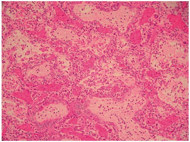
Figure 1. Lung histopathology of ACD/MPV.
HE staining, × 100. The lobular architecture is altered with enlarged simplified and poorly subdivided alveoli. Small pulmonary arteries adjacent to bronchioles show mildly increased amounts of smooth muscle within their walls; there are abnormally positioned markedly dilated thin-walled veins adjacent to these small pulmonary arteries. Capillaries cannot be clearly delineated at this magnification, but they are deficient in numbers in the thickened alveolar walls, where there are larger centrally located dilated thin-walled vascular channels. Clusters of neutrophils are evident in some airspaces (perinatal pneumonia).

