
Fig. 2.
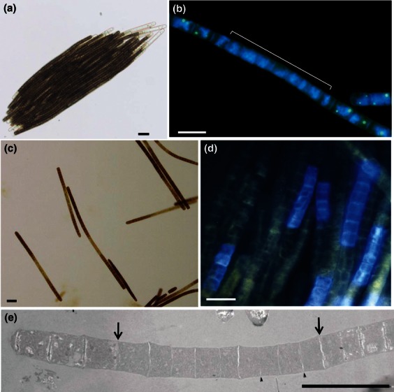
Morphological characteristics of Trichodesmium trichomes, with emphasis on cell differentiation and their nitrogenase containing cell type, the diazocytes. (a) A light micrograph depicting a dark pigmented colony consisting of longitudinally arranged trichomes of a newly isolated strain, T. erythraeum TNZ0801. Scale bar, 25 μm. (b) The DNA distribution in cells of a Trichodesmium IMS101 trichome visualized after staining with the dye 4′,6-diamidino-2-phenylindole (DAPI), fluorescing blue. Note the DNA presence in all cells, the centrally located diazocyte-like zone (marked) being recognized as they are devoid of the yellow/green fluorescent granules representing polyphosphate storage. Scale bar, 20 μm. (c) Trichomes of Trichodesmium IMS101 stained with Lugol's solution. Note several lighter-stained central diazocyte zones, in which catabolic carbon metabolism has degraded the Lugol-stainable stored carbon supplies. Scale bar 20 μm. (d) Fluorescence in situ immunolocalization of NifH into groups of adjacent cells, diazocytes, in central areas of intact trichomes of Trichodesmium IMS101. The NifH protein is detected as a blue fluorescence due to a secondary anti-NifH-antibody coupled to a blue-fluorescing chromophore. Scale bar, 10 μm. (e) Transmission electron micrograph depicting a longitudinally sectioned trichome of Trichodesmium IMS101. Note the more homogenous zone of cells, representing diazocytes between the arrows. Arrowheads point to ongoing cell division (the formation of division septa) in two of the diazocytes. Scale bar, 20 μm.