Figure 7.

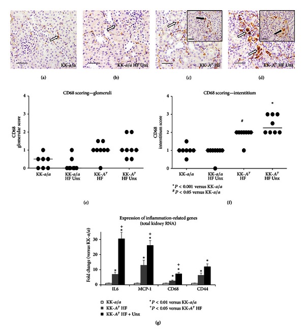
Increased inflammation in KK-A y mice.Macrophage infiltration was evaluated by CD68 immunohistochemistry. KK-a/a (a) and KK-a/a + Unx (b) mice had scattered CD68+macrophages in the interstitium (white arrows) and rare CD68+cells in the glomeruli. KK-A y (c) and KK-A y + Unx (d) mice showed increased CD68+macrophage infiltration in the interstitium (white arrows) and occasional CD68+cells in glomeruli (inset, black arrows). Scale bars represent 50 μm. Semiquantitative scoring of CD68+ cells revealed a modest increase in positively stained cells within glomeruli of KK-A y relative to the KK-a/a controls (e), additionally a statistically significant increase in interstitial CD68+ macrophages in both groups, with a slight enhancement in the KK-A y + Unx group (f). Horizontal bars indicate group median score and asterisk represents P value <0.001. (g) Gene expression was assayed by qRT-PCR using total kidney RNA from 26 week old mice KK-A y, uninephrectomized KK-A y, and control KK-a/a animals. qRT-PCR revealed increased expression of IL-6, MCP-1, CD68, and CD44 were significantly upregulated in KK-A y versus KK-a/a (P < 0.01). Uninephrectomy further exacerbated IL-6, MCP-1, and CD68 mRNA upregulation versus KK-A y (P < 0.05).
