
Figure 7.
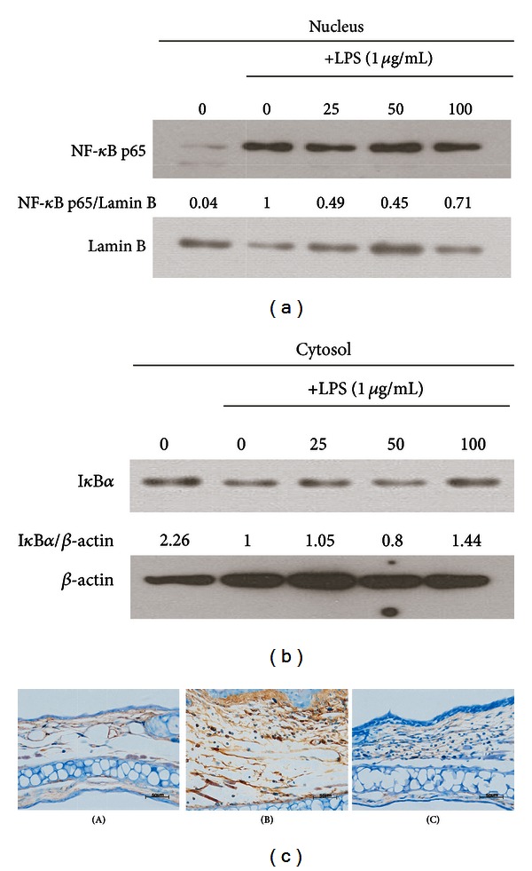
Effects of RV extract on the Iκ-B and NF-κB in LPS-stimulated RAW264.7 macrophages. (a, b) Cells were incubated for 30 min with 1 μg/mL of LPS in the absence or presence of RV extract. RV extract was added 1 h before the incubation with LPS. The protein expression of Iκ-B and NF-κB was analyzed by Western blot analysis and quantified by densitometric analysis. (c) The immunohistochemical analysis was used to monitor the protein expression of NFκB in the ear sections. Scale bar = 50 μm. Original magnification, 400x. (A) Control, (B) RV(−), (C) RV(+). Data are represented as mean ± standard error (S.E.) of three independent experiments. One-way ANOVA was used for comparisons of multiple group means followed by Dunnett's t-test (# P < 0.001 versus control; *P < 0.01, **P < 0.05, ***P < 0.001 versus LPS-stimulated control).