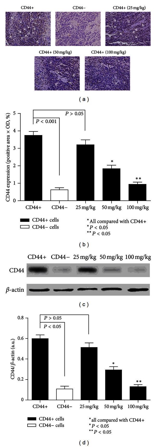
Figure 6.

(a) Immunohistochemical staining of CD44. CD44 was highly expressed in the cancer cell membrane (original magnification 400x, positive areas are indicated by white arrows). (b) CD44 expression in each of the treatment groups (N = 6 in each groups). CD44 was highly expressed in model CD44+ group compared to the rest of the groups and was rarely detected in model CD44− group. The results also showed that not all of the cells in the CD44+ group were CD44 positive. β-Elemene inhibited the expression of CD44 in a dose-dependent manner. Statistically significant differences in expression of CD44 were also detected in the 50 mg/kg and 100 mg/kg groups (P < 0.05). (c)-(d) Western blot results of CD44 expression in model CD44+ group, model CD44− group, and all the β-elemene-treated CD44+ groups. We detected a corresponding variation of CD44 in these groups like the aforementioned IHC results of it.
