Figure 1.

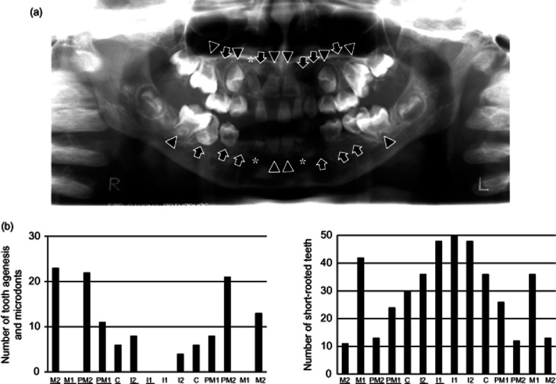
Emergence of chemotherapy-induced tooth formation anomalies. (a) Orthopantomograph of a case of chemotherapy-induced crown formation anomalies. The subject was treated with high-dose chemotherapy (HDC) at 1 year of age and the oral examination here was at 12 years of age. All permanent teeth except the third molars have anomalies: the bilateral upper lateral incisors, upper left canine, bilateral upper second premolars, bilateral lower canines, and bilateral lower first and second premolars display tooth agenesis (TA, arrows); the upper right canine and bilateral lower lateral incisors microdonts (MO, asterisks); teeth not subject to TA/MO short-rooted teeth (SR, arrowheads). (b) Permanent teeth with chemotherapy-induced anomalies. Central and lateral incisors, canines, first and second premolars, and first and second molars are expressed as I1 and I2, C, PM1 and PM2, and M1 and M2. Upper teeth are underlined. The highest incidence was in the second premolars and second molars with tooth agenesis or microdonts, and in the incisors with short-rooted teeth. Tooth agenesis or microdonts did not occur in the upper and lower first molars and central incisors.
