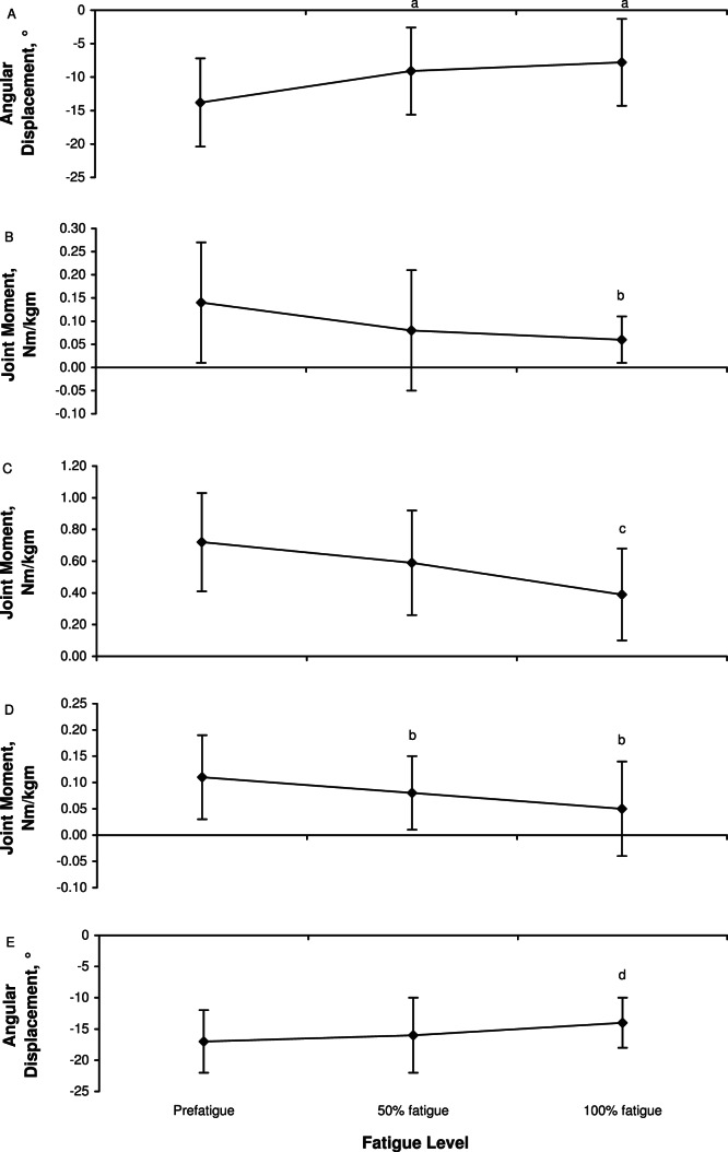
Figure 1.
Changes in lower extremity biomechanics throughout the fatiguing protocol for hip-abduction angles at initial contact, A, hip-abduction moment at initial contact, B, hip-abduction moment at peak stance, C, knee-abduction moment at initial contact, D, and knee-flexion angle at initial contact, E. a Indicates less abducted than at prefatigue. b Indicates less than at prefatigue. c Indicates less than at prefatigue and 50% fatigue. d Indicates less knee flexion than at prefatigue and 50% fatigue. These variables presented a progressive deterioration from prefatigue to 100% fatigue.

