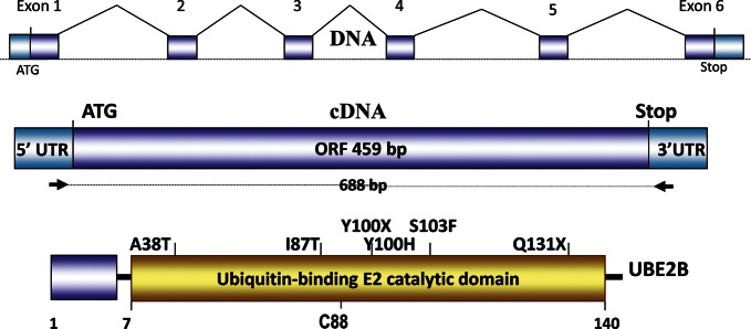
Figure 1.

UBE2B gene, cDNA and point alterations identified in severe oligozoospermic patients. Schematic structure of the UBE2B gene, cDNA amplification strategy and single nucleotide mutations in the gene are shown. The upper diagram depicts the normal splicing pattern. The positions of the single nucleotide mutations and variants in the UBE2B protein are shown. The position of the cysteine residue (C) is shown at the bottom. The UBC E2 domain is predicted at 7–140 amino acids. The protein structure is based on Interproscan, Prosite, Pfam and Blocks algorithm predictions.
