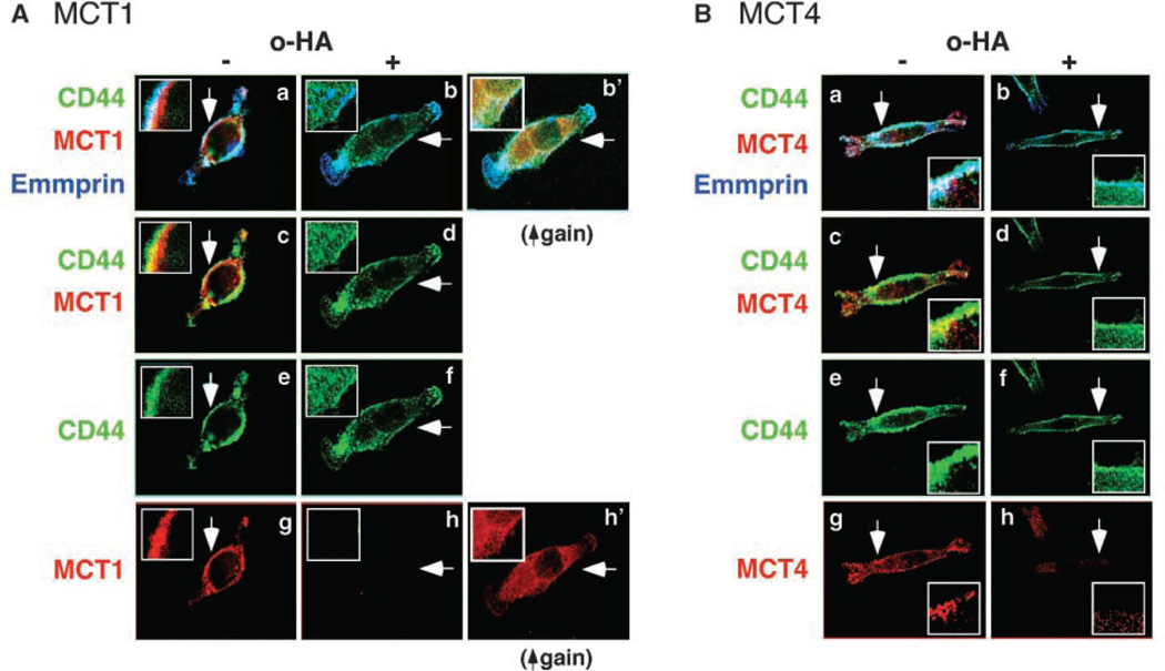
Figure 3.
CD44 and emmprin colocalize with MCT1 and MCT4 at the plasma membrane in a hyaluronan-dependent manner. A, MCT1; B, MCT4. MB231 cells were treated with and without 100 µg/mL hyaluronan oligomers for 1 h. After fixation, permeabilization, and blocking of nonspecific binding, the cells were immunolabeled for CD44 (green), MCT1 or MCT4 (red), and emmprin (blue) and visualized by confocal microscopy at a Z plane corresponding to the approximate center of the cell. Arrows, areas shown at higher magnification in the insets. a, b, and b’, triple labeling for all three components; colocalization appears as a white signal. Triple colocalization is clearly seen in the plasma membrane of the untreated cells but not in the membrane of oligomer-treated cells. Increased intracellular staining for CD44 is seen in the oligomer-treated cells in b. b’ in A is the same as in b, but is shown at higher gain than in a and b, to illustrate internalization of MCT1, as well as CD44; increased gain is necessary for clear visualization because the MCT becomes highly dispersed in the cytoplasm after oligomer treatment. Similar oligomer-induced internalization was observed for MCT4 (not shown). Emmprin seems to remain at the cell surface after hyaluronan oligomer treatment. c and d, double staining for CD44 and MCT1 (A) or MCT4 (B), further illustrating their colocalization (yellow signal in c) and hyaluronan oligomer-induced internalization of CD44 (d). e and f, CD44 only; g and h, the MCTs only. Increased gain revealed intracellular dispersion of MCT1 (A, h’) and MCT4 (not shown). The figure is representative of three or more independent experiments.

