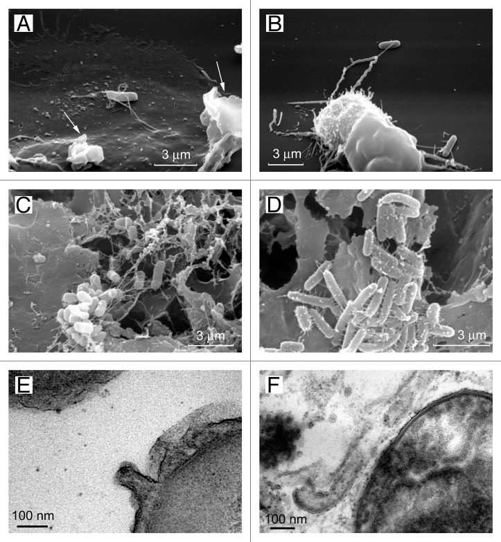
Figure 5. Cytonemes of bacteria. The scanning electron microscopy images show Salmonella enterica serovar Typhimurium attached to the surface of the control neutrophils (A) or to the TVEs of BPB-treated neutrophils (B) through cytonemes. Salmonella of the virulent C53 strain (C) and the non-flagellated SJW880 strain (D) were interconnected through cytonemes in biofilms grown on the surface of gallstones. Transmission electron microscopy images of 60-nm membrane tubules derived from the outer membrane of the bacteria (E and F).34

An official website of the United States government
Here's how you know
Official websites use .gov
A
.gov website belongs to an official
government organization in the United States.
Secure .gov websites use HTTPS
A lock (
) or https:// means you've safely
connected to the .gov website. Share sensitive
information only on official, secure websites.
