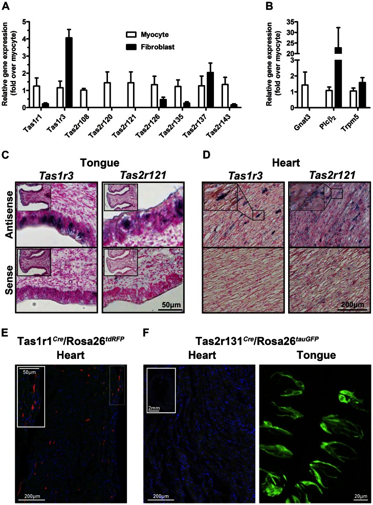
Figure 3. Taste GPCRs are localized in isolated cardiac cells.
A Taste receptors were detected in cultured neonatal cardiomyocytes and fibroblasts by RT-qPCR (mean±SEM, n = 4, normalized for Gapdh and expressed relative to myocytes). B The taste receptor signal transduction genes, including G protein (Gnat3), second messenger (PLCβ2) and channel (Trpm5) are also expressed in myocytes and fibroblasts (mean±SEM, n = 4, normalized for Gapdh and expressed relative to myocytes). C In situ hybridization using digoxigenin-labeled cRNA probes specific for Tas1r3 (left panels) and Tas2r121 (right panels) show expression in the taste buds of the circumvallate papillae (inset shows low magnification) and D in heart tissue (inset shows higher magnification of boxed region). Specific labeling (blue/black) is observed using antisense probes, but not sense probes. E Tas1r1 is expressed in mouse ventricular tissue from Tas1r1Cre/Rosa26tdRFP mice, where red fluorescent cells report activity of the Tas1r1 promoter. F In contrast, in heart sections from the Tas2r131Cre/Rosa26tauGFP reporter mouse line, there are no fluorescently-labeled myocytes (inset shows low magnification view of heart). Tas2r131–expressing cells are clearly labeled in the circumvallate papillae as control. Scale bars are as indicated.

