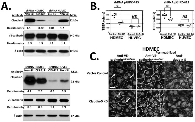
Figure 6.
ShRNA knockdown of claudin-5 in HDMECs and HUVECs. (A) Top: Immunoblot analysis of claudin-5 expression in HDMECs (left) and HUVECs (right) transduced with non-silencing lentivirus vector control (Non-Sil) or with claudin-5 shRNA clone V2LHS_171415 (CL5 KD; top panel) or with a second claudin-5 shRNA, clone V2LHS_171412 (bottom panel) at day 4 post-visual confluence. Numbers indicate densitometric analyses of the immunoblot data normalized to expression of β-actin. (B) TEER levels in HDMECs or HUVECs stably transduced with shRNA pGIPZ clone V2LHS_171415 (pGIPZ-415) or clone V2LHS_171412 (pGIPZ-412) vs. non-silencing (Non-Sil) shRNA negative control-transduced ECs on day 3 post-confluence. Knockdown of claudin-5 in HDMECs significantly reduced TEER (p < 0.0001 and p < 0.005 by two-tailed t test for GIPZ-415 and GIPZ412, respectively) whereas no significant TEER differences are observed in HUVECs transduced with either shRNA. TEER values shown are means from multiple experiments with pGIPZ-415 (n = 5, 3) and with pGIPZ-412 (n= 5, 5). (C) Confocal fluorescence optical z-sections of vector control (upper panels) vs. claudin-5 knockdown HDMEC (lower panels) monolayers at day 4 post-visual confluence immunostained with mouse mAb BV6 to VE-cadherin extracellular epitopes in non-permeabilized cells (arrows indicate differential antibody accessibility) and goat anti-VE-cadherin antibody to intracellular epitopes or rabbit anti-claudin-5 antibody in permeabilized cells. Scale bar = 15 μm. One of three experiments with similar results.

