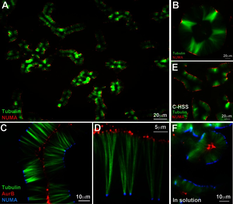
FIGURE 1:
Pineapple assembly. (A) Typical field 1 h after addition of 5 μM Taxol and 5% DMSO to meiotic HSS containing tubulin (green) and NUMA (red) probes; 20× wide-field imaging. Note formation of similar assemblies across the whole field, comprising paired lines of microtubule bundles with NUMA aligned on the outside. (B) Round assemblies with open centers resembled a slice of canned pineapple (∼2 h of assembly, 20× wide field). Tubulin (green), NuMA (red). (C) A 40× confocal image at 90 min showing localization of Aurora B (red) on the inside and NUMA (blue) on the outside. Note global alignment of both types of foci on curved planes, which appear as lines in optical sections. (D) A 100× confocal image taken at 90 min showing one side of an open assembly. NUMA (blue) was on the outside as usual. Both NUMA and Aurora B (red) accumulate at foci. Aurora B foci are smaller and more numerous. (E) Pineapple assembly in C-HSS, which is completely free of organelles; 120 min, 20× wide field. Tubulin (green), NUMA (red). (F) Pineapple assembly in solution. This image was taken by assembly in solution for 1 h, followed by 20× dilution into a Taxol-containing buffer without DMSO, squashing between a slide and coverslip, and imaging within ∼5 min (40× confocal). Other examples are shown in Figure 4. NUMA (blue), Aurora B (red), tubulin (green).

