
FIGURE 2:
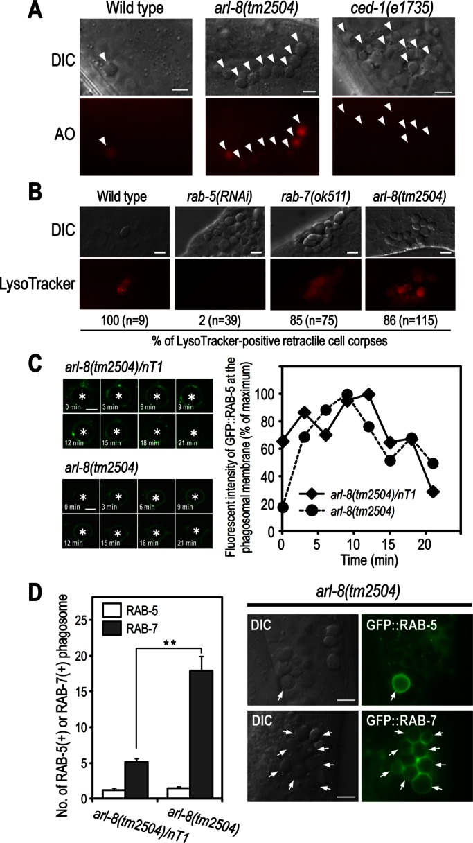
Phagosomes are arrested at an RAB-7–positive stage in arl-8(tm2504) mutants. (A) AO staining of germ cell corpses in the indicated animals. Germ cell corpses are marked with arrowheads. Most cell corpses were stained by AO in arl-8(tm2504) animals, whereas corpses were not stained in ced-1(e1735) animals in which cell corpses are not phagocytosed by sheath cells. Bars, 5 μm. (B) LysoTracker staining of germ cell corpses and quantitation of LysoTracker-positive cell corpses in the indicated animals. n, number of cell corpses scored. Bars, 5 μm. (C) Time-lapse images of GFP::RAB-5 expression at the phagosome (left) and quantitation of the fluorescence intensity (right) in arl-8(tm2504)/nT1 (as a control) and arl-8(tm2504) animals. Asterisks indicate germ cell corpses. Bars, 2.5 μm. (D) Quantitation of RAB-5–positive and RAB-7–positive phagosomes in the germline of the indicated animals (left). Fifteen animals were scored for each strain, and data are expressed as mean ± SEM. **p < 0.01 by Student's t test. Nomarski and fluorescence images of arl-8(tm2504) animals expressing GFP::RAB-5 or GFP::RAB-7 (right). Arrows indicate the germ cell corpses positive for the corresponding GFP-marker proteins. Bars, 5 μm.