
FIGURE 6:
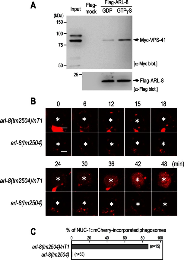
ARL-8 physically interacts with VPS-41 and is required for phagosome–lysosome fusion. (A) Pull-down assay using Flag-ARL-8 and Myc-VPS-41. Purified Flag-ARL-8 proteins preloaded with either GDP or GTPγS were incubated with HEK293T cell lysates containing Myc-VPS-41 and immunoprecipitated using anti-Flag M2 agarose. The precipitated fractions were then subjected to Western blot analysis using the indicated antibodies. (B) Time-lapse images of NUC-1::mCherry signal in the gonad of arl-8(tm2504)/nT1 and arl-8(tm2504) animals. Asterisks indicate germ cell corpses. Here 0 min represents the point at which GFP::RAB-5 (not shown) was first detected on phagosomal membranes. Bars, 2.5 μm. (C) Quantitation of fusion events in the indicated animals. Data are expressed as the ratio of cell corpses that incorporated NUC-1::mCherry within 1 h after their appearance. n, number of cell corpses scored.