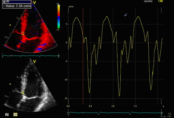
Figure 1.

How the peak systolic velocity (PSV) information is acquired. The region of interest (ROI) is manually placed in the basal segments of the left ventricle and the corresponding tissue velocity is showed in the graph to the right where you can register the PSV guided by the ECG below in the picture. The time is on the x-axis and velocity in cm/s on the y-axis.
