
Figure 3.
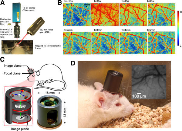
(A) Setup of the experimental LSCI [72]. (B) Images displaying the vascular responses during and after electrical stimulation of peripheral trigeminal nerve fibers [28]. (C) A schematic drawing of the integrated imaging microscope shown that the incident and reflected light paths in blue and green, respectively; and a photograph of the assembled device [73]. (D) This integrated imaging microscope can be used for untethered cortical imaging in freely-moving animals [73].