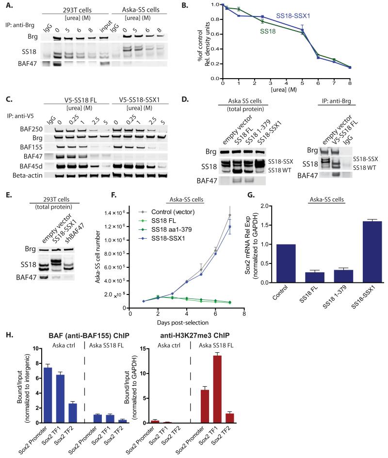
Figure 6. Reversible integration, gene expression and occupancy by SS18 and SS18-SSX containing mSWI/SNF (BAF) complexes.
(A) Denaturation studies using 0-8M urea with subsequent immunoblot analysis for SS18 in 293T cells and SS18-SSX in Aska-SS cells. See also Figure S6.
(B) Quantitative densitometry of SS18 or SS18-SSX1 protein immunoblots from n=3 experimental replicates of urea denaturation 0<[urea]<8M. Y-axis: band quantitation/ untreated control. Error bars= s.d.
(C) IP using anti-V5 antibody in urea treated nuclear extracts isolated from 293T fibroblasts infected with either V5-SS18 or V5-SS18-SSX with immunoblotting for BAF complex components.
(D) Left, Immunoblot analysis on total protein isolated from Aska-SS cells with either SS18 or SS18 1-379 or SS18-SSX1 introduced via LV. Right, anti-Brg IP of complexes in either empty vector or V5-SS18FL treated conditions.
(E) Introduction of SS18-SSX1 and shBAF47 into 293T cells with subsequent immunoblot analysis on total protein.
(F) Cell proliferation analyses of Aska-SS cells infected with control vector, SS18, SS18 1-379, and SS18-SSX. Error bars= s.d.
(G) Sox2 mRNA relative expression (normalized to GAPDH) 10 days post infection with LV containing either control shScramble or overexpression of SS18, SS18 1-379, or SS18-SSX. Error bars= s.d.
(H) Left, anti-BAF155 ChIP on Aska-SS cells treated with either empty vector or SS18 FL, with subsequent qPCR for regions at the human Sox2 promoter and two Sox2 transcription factor (TF) binding sites within the exon. Right, anti-H3K27me3 ChIP at Sox2 locus in Aska-SS control treated and SS18 FL-treated cells. Error bars= s.d.

