
Figure 1.
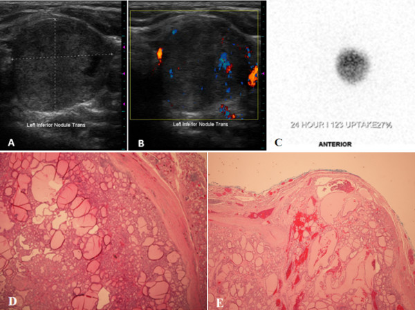
Imaging and histologic features of the hot nodule present in the case report subject. (A) Ultrasonography of the left thyroid lobe, demonstrating a 2.7 cm, predominantly solid, and isoechoic nodule. (B) Color Doppler evaluation reveals blood flow within the rim of the nodule and intraparenchymally. (C) 123I thyroid scintigram depicts a round left-sided focus of iodine uptake with suppression in the remainder of the gland, consistent with an autonomously-functioning thyroid nodule. (D) Histological evaluation reveals that the lesion is solitary, circumscribed and encapsulated. The follicular proliferation is surrounded by a rather thick fibrous capsule. The lesion demonstrates a predominant follicular pattern of growth without papillary cytologic features (hematoxylin-eosin stain; original magnification × 4). (E) A focal area is identified where the tumor invades through and into the fibrous capsule (hematoxylin-eosin stain; original magnification × 2).