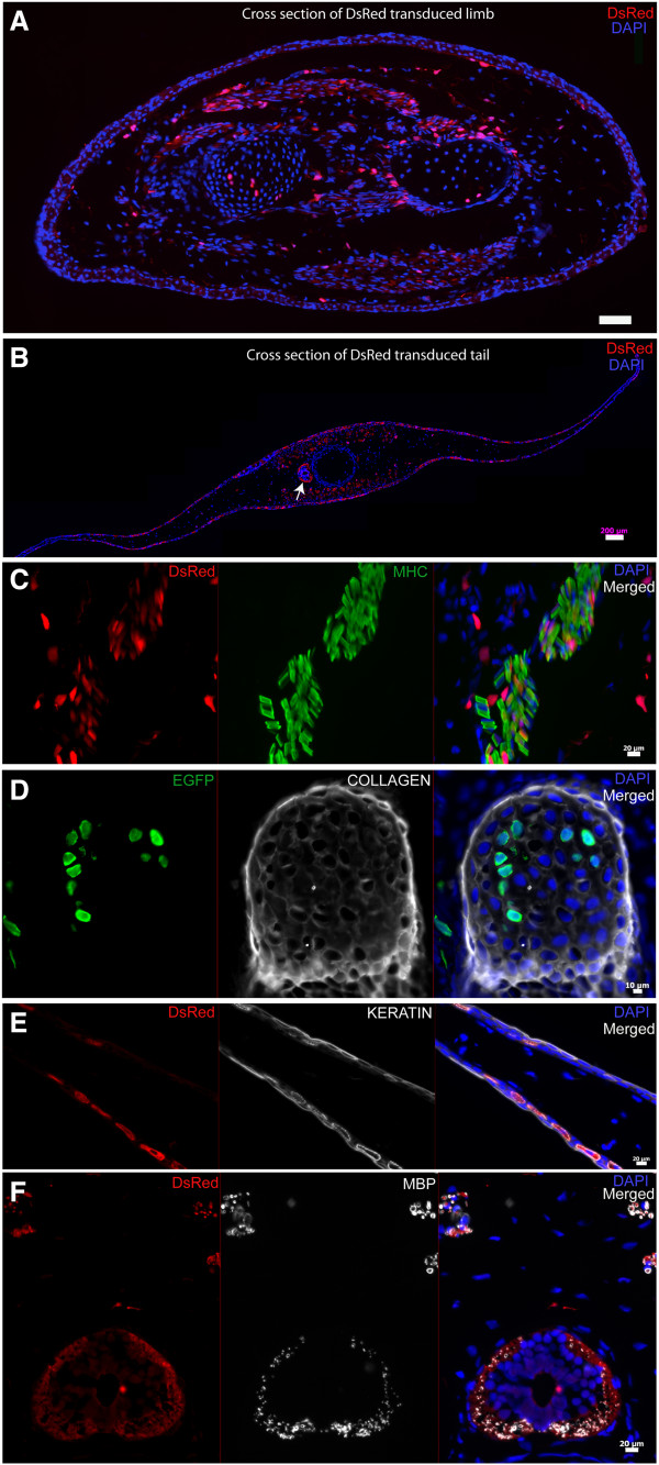
Figure 4.
Foamy virus vector transduce muscle, cartilage, dermis, Schwann cells in the regenerating axolotl limb and tail. A. Cross section of limb shown in Figure 3C (transduced with FV-DsRed) showing wide spread DsRed expression. B. Cross section of tail shown in Figure 3D (transduced with FV-DsRed) showing wide spread DsRed expression. Arrow points to the spinal cord. C. Immunofluorescence staining for muscle specific myosin heavy chain (MHC) in limb (transduced with FV-DsRed) showing endogenous DsRed expression (left), MHC staining (middle) and merged of the two images is shown on the right. (DAPI=blue). D. Immunofluorescence staining for collagen in limb (transduced with FV-EGFP) endogenous EGFP expression is on the left, collagen staining (middle) and merged of the two images is shown on the right. (DAPI=blue). E. Immunofluorescence staining for keratin in tail (transduced with FV-DsRed). The image shown is a blow up of the tail fin region showing excellent colocalization of endogenous DsRed expression (left) with keratin immunostaining (middle). F. Immunofluorescence staining for myelin basic protein (MBP) in tail (transduced with FV-DsRed). The image depicted here shows the spinal cord and associated peripheral nerve tracts showing endogenous DsRed expression (left), MBP staining (middle) and merged of the two images on the right. Scale bars: A: 100 μm, B: 200 μm, C,E,F: 20 μm, D: 10 μm.

