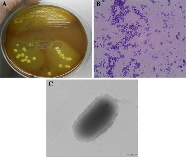
Figure 1.
Colonial morphology, gram staining and transmission electron microscopic image of the CF clinical isolate Microbacterium yannicii PS01. A. CF clinical isolate Microbacterium yannicii PS01 was grown on Columbia colistin-nalidixic acid agar with 5% sheep blood (bioMérieux) at 37°C with 5% CO2. The colony appeared as yellow, round and smooth. B. Gram staining picture of the gram-positive coccobacilli CF clinical isolate “CF Microbacterium yannicii PS01” viewed at 100X magnification. C. Transmission electron microscopy image of M. yannicii strain PS01, using a Morgani 268D (Philips) at an operating voltage of 60kV. The scale bar represents 900 nm.

