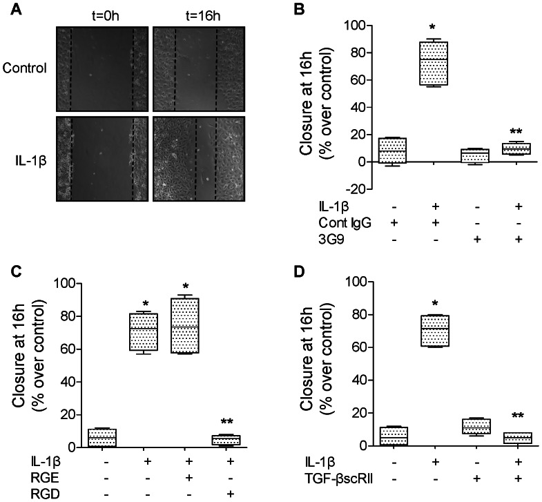
Figure 4. IL-1β increases wound closure via αvβ6 integrin and TGF-β1 in primary rat ATII cell monolayers.
(A) IL-1β increases the rate of wound closure of primary rat ATII cell monolayers. IL-1β (10 ng/ml) or its vehicle was added to the monolayers after the scratch. Phase contrast microscopy (20X magnification) immediately after wounding (left panels, t = 0 h) and after 16 h (right panels t = 16 h). Scale bar: 100 µm. (B) A β6 blocking antibody (3G9) prevents IL-1β-dependent increase in rate of wound closure of a primary rat ATII cell monolayer. IL-1β (10 ng/ml). A β6 blocking antibody or its isotype control antibody was added to the monolayers after the scratch. (C) RGD peptides prevent IL-1β-dependent increase in rate of wound closure of a primary rat ATII cell monolayers. IL-1β (10 ng/ml) and RGE or RGD peptide were added to the monolayers after the scratch. (D) A TGF-β1 soluble receptor (TGF-βscRII) prevents IL-1β-dependent increase in rate of wound closure of primary rat ATII cell monolayers. IL-1β (10 ng/ml) and/or TGF-βscRII or their respective vehicles were added to the monolayers after the scratch. Degree of wound closure is expressed as percent of control 16 h after wounding. *p<0.05 from monolayers exposed to IL-1β vehicle. **p<0.05 from monolayers exposed to IL-1β.

