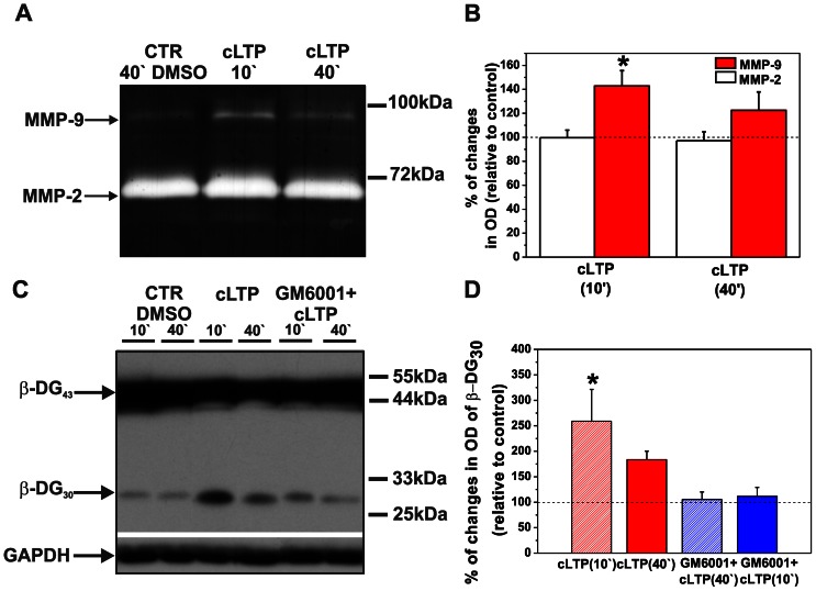
Figure 1. cLTP increases endogenous MMP-9 but not MMP-2 activity and increases β-DG cleavage by MMP-9.
(A) Cortical neurons were exposed to the F/R/P mixture for 10 and 40 min, and gelatinase activity (MMP-9 and MMP-2) was then assayed. A representative gel zymogram shows enhanced MMP-9 activity compared with controls at 10 min, which was followed by a decrease at 40 min of cLTP stimulation. Notice that the enzymatic activity of MMP-2 did not change during cLTP. (B) Column bars present the quantification (mean ± SEM) of MMP-9 and MMP-2 activity 10 and 40 min after F/R/P treatment relative to control (DMSO-treated cells). Student’s t-test revealed a significant increase in the level of total amount of MMP-9 at 10 min. The level of MMP-9 did not significantly differ from controls after 40 min of cLTP. Student’s t-test did not reveal significant changes in the total amount of MMP-2 compared to control after 10 and 40 min of cLTP. (C) Cortical neurons were exposed to the F/R/P mixture, and β-DG cleavage was then tested by Western blot. A representative Western blot shows enhanced proteolysis of β-DG 10 min after stimulation compared with controls. Forty minutes of cLTP stimulation decreased the level of the cleaved form of β-DG. Blocking MMP activity with GM6001 led to a further decline of β-DG cleavage. Control cells were incubated with DMSO. Glyceraldehyde-3-phosphate dehydrogenase (GAPDH) served as the loading control. (D) Column bars indicate results of relative densitometric analysis of β-DG cleavage 10 and 40 min after F/R/P treatment relative to control (DMSO-treated cells). Students t-test showed statistically significant increase in cleaved form β-DG at 10 in of cLTP. Students t-test failed to reach significance for β-DG cleavage at 40 min of F/R/P exposure and for cLTP stimulation in the presence of MMP inhibitor, GM6001. Results are mean ± SEM, n = 3, *p<0.05.

