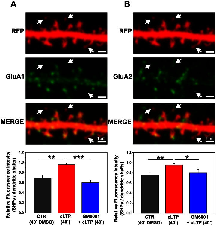
Figure 5. cLTP drives GluA1- and GluA2-containing AMPARs into spines that carry SHPs in an MMP-dependent manner.
A short segment of a neuronal dendrite labeled with anti-GluA1 (A) and anti-GluA2 (B) antibodies is shown at 40 min of cLTP. Arrows indicate spines with SHPs. Bar plots show the quantification of fluorescence readouts from immunostaining against GluA1 (A) and GluA2 (B) subunits within spines that exhibited SHPs vs. dendritic shafts. (A). Students t-test revealed significant differences for GluA1-containing AMPAR content at spines with processes compared with control (DMSO-treated cells) after 40 min cLTP. Blocking MMP activity with GM6001 abolished GluA1-containing AMPARs surface delivery into spines with protrusions. (B) Forty minutes of cLTP induced the surface delivery of GluA2-containing AMPARs into spines with SHPs in an MMP-dependent manner. Students t-test showed significant differences for cLTP-stimulated cells compared with control and GM6001-pretreated groups. Results are mean ± SEM, n = 6, *p<0.05, **p<0.01, ***p<0.001.

