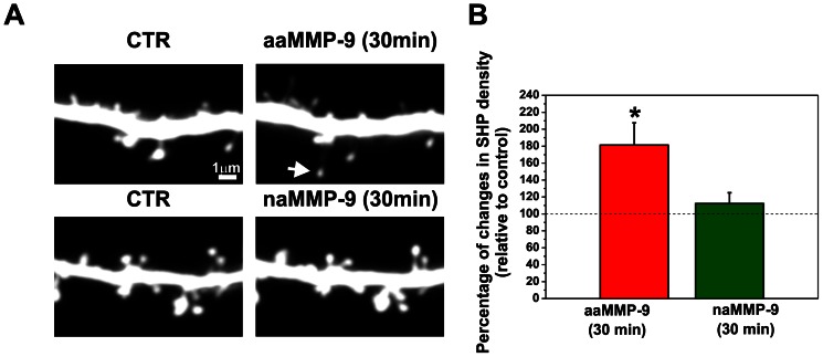
Figure 6. Enzymatic activity of MMP-9 is required for SHP development.
(A) A segment of a secondary apical dendrite from a pyramidal neuron that expressed RFP was imaged with a confocal microscope (CTR) and then treated with autoactive MMP-9 (aaMMP-9) for 30 min. The white arrow shows a SHP formed after 30 min treatment with active MMP-9. Treatment with inactive MMP-9 did not lead to the formation of SHPs. (B) Bar plot shows the quantification of new SHPs per µm of dendrite. Students t-test revealed statistically significant increase in newly formed SHPs after treatment with active MMP-9. Incubation with inactive MMP-9 had no effect on the formation of SHPs Results are mean ± SEM, *p<0.05.

