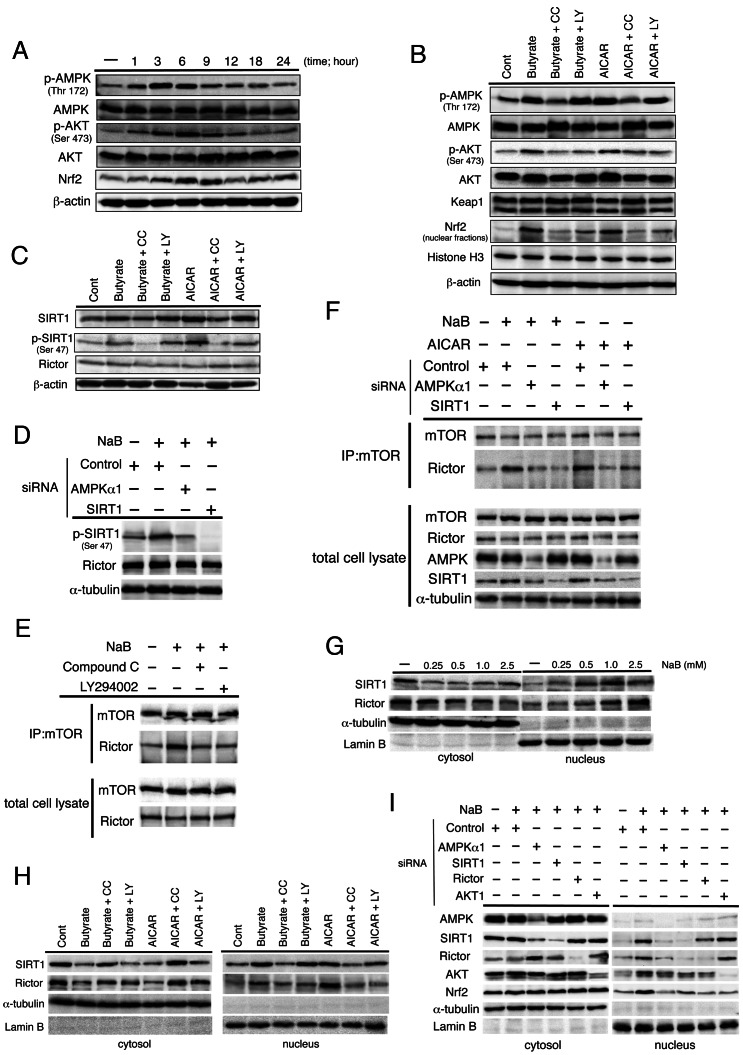
Figure 6. Nrf2 expression is regulated by AMPK and AKT activation and mTORC2 modification underlying NaB treatment.
(A) Serum-starved HepG2 cells were treated with 1.5 mM NaB for the indicated time periods. Western blot analysis was performed with the indicated antibodies. β-actin expression was used as the loading control. (B, C) Serum-starved cells were pretreated for 1 h with AMPK agonist compound C (CC; 20 µM) or the PI3K-specific inhibitor LY294002 (LY; 25 µM) and then incubated with 1.5 mM NaB or 1 mM AMPK activator AICAR for 6 h. Western blot analysis was performed with the indicated antibodies. (D) Cells were transfected with indicated siRNAs for 48 h and then incubated with 1.5 mM NaB for 6 h under serum-starved conditions. Western blot analysis was performed with the indicated antibodies. α-tubulin expression was used as the loading control. (E) Serum-starved cells were pretreated for 1 h with 20 µM CC or 25 µM LY and then incubated with 1.5 mM NaB for 6 h. Cell lysates and mTOR immunoprecipitates (IPs) prepared from the total cell lysates were analyzed by western blotting for the levels of mTOR and rictor. (F) Cells were transfected with the indicated siRNAs for 48 h and then incubated with 1.5 mM NaB or 1 mM AICAR for 6 h under serum-starved conditions. Cell lysates and mTOR immunoprecipitates (IPs) prepared from the total cell lysates were analyzed by western blotting for the levels of mTOR and rictor. (G) Nuclear accumulation of SIRT1 or rictor was examined by western blot analysis. Cells were treated with NaB for 6 h at the indicated concentration. Anti-α-tubulin and anti-lamin B antibodies were used as markers for the cytoplasmic and nuclear extracts, respectively. (H) Serum-starved cells were pretreated for 1 h with 20 µM CC or 25 µM LY and then incubated with 1.5 mM NaB for 6 h. Nuclear accumulation of SIRT1 or rictor was examined by western blot analysis. α-tubulin and lamin B were evaluated for expression levels as markers for the cytoplasmic and nuclear extracts, respectively. (I) Cells were transfected with indicated siRNAs for 48 h and then incubated with 1.5 mM NaB for 6 h under serum-starved conditions. Western blot analysis was performed with the indicated antibodies. α-tubulin and lamin B expression were used as the loading control of cytoplasmic and nuclear extract proteins, respectively.

