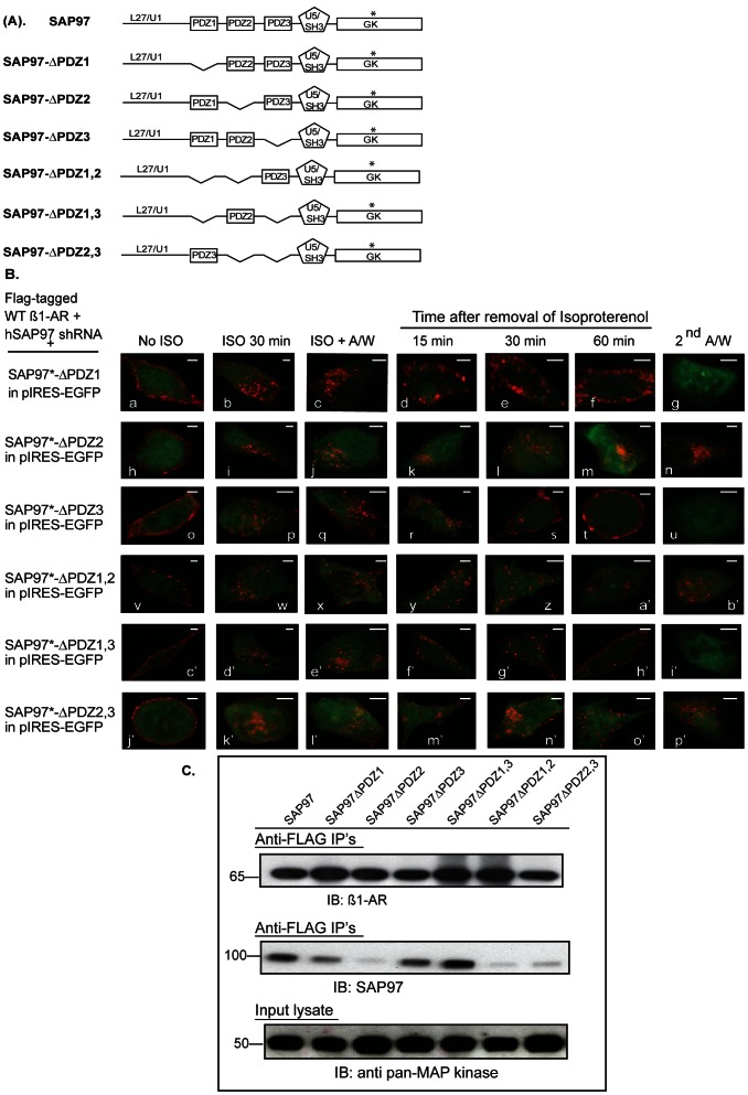
Figure 3. Mapping of the role of SAP97 PDZ domains in binding to and in recycling of the ß1-AR.
A, schematic diagram of SAP97 and deletion constructs used in panels B and C. B, cells stably expressing FLAG WT ß1-AR and hSAP97 shRNA were transiently transfected with rSAP97 or its deletion constructs in pIRES-EGFP II that are shown in panel 3A. The internalization and recycling assays were carried out as described in the legend of Fig. 2. Each scale bar represents 5 µm. C, HEK-293 cells expressing hSAP97 shRNA and FLAG-tagged ß1-AR were transfected with pIRES-EGFP harboring SAP97 or its various PDZ deletion mutants shown in panel A above. Equal amounts of cell lysates (input) were incubated with ∼8 µL of anti-FLAG M2 IgG resin overnight. Then the beads were washed and eluted into 50 µl of 2X-Laemmli sample buffer. The top panel shows Western blot analysis of 10% of each FLAG IP that were probed with anti-human ß1-AR IgG (IB) to index ß1-AR IP’s. The middle panel shows Western blot analysis of 80% of each FLAG IP that were probed with anti-SAP97 IgG (IB) to index SAP97 Co-IPs. To normalize for input protein levels, ∼4% of each cell lysate were subjected to Western blotting (IB) and probed with the anti-pan MAP kinase antibody.

